


DEXA Scan
A DEXA bone density scan, also known as 'dual-energy X-ray absorptiometry,' is a painless, non-invasive imaging diagnostic technique used to measure your bone mineral density and the level of bone loss.
The DEXA scan results assist orthopedics in confirming the presence or risk of osteoporosis and fractures.
The DEXA bone density test can help you figure out how strong your bones are and how vulnerable you are to fractures and other bone problems.
The hips and spine, two of the body's most vulnerable skeletal systems, are the focus of this bone imaging examination.
The DEXA bone scan is an outpatient procedure that takes around 10 to 20 minutes and allows you to return home the same day.
Because he uses two X-ray beams, a high energy beam and a low energy beam, his bone scanning approach is far more efficient than a standard X-ray.
T-score refers to the findings of a DEXA bone scan. The bone health of a 30-year-old is used as a benchmark in this grading system because the bones are at their strongest at this age.
Procedure for a DEXA Scan
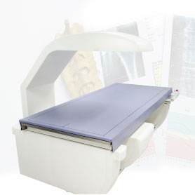
You must lie flat on your back on a flat and comfortable padded surface for the DEXA bone density scan. To produce accurate images, a wedge device can be utilised to modify the posture of your spine and hips.
During the exam, you must remain still, and the radiologist may ask you to hold your breath for a few seconds if necessary.
The X-ray instrument that takes the images for evaluation is housed in a moving arm of the DEXA scanner. The arm gently glides across the body, capturing high-resolution photos of the bones.


DEXA Scan is a bone density test scan that is used to determine the health of the body.
As we age, our bones also become weak and DEXA Scan becomes important so that we can prevent major injuries.
At Star Imaging and Path Lab is the best DEXA scan near me in Delhi NCR which provide 100% accurate report to the patient.

