


Breast radiation image photography utilizes various technologies to examine breast tissue for abnormalities that could indicate breast cancer or other issues. Understanding the different modalities used can help in choosing the right exam.
Mammography
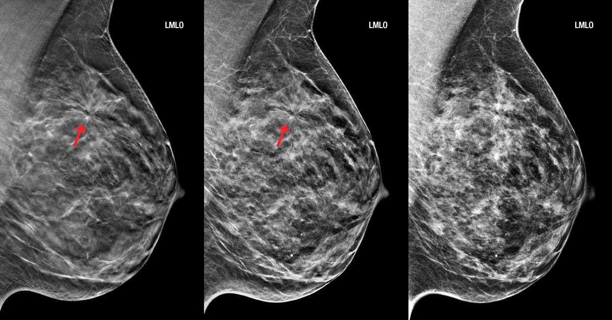
Mammography is considered the gold standard breast cancer screening tool. A low-dose x-ray of the breast is used to detect subtle changes in breast tissue up to two years before a physical exam. Digital mammograms can find 30-40% more invasive Breast Imaging than film mammograms and are now the standard of care in the U.S. Mammograms are best at finding microcalcifications and masses but have limitations in detecting tumors in women under 50 with dense breast tissue.
Breast Ultrasound
An ultrasound uses high-frequency sound waves to create images of breast tissue. It is helpful in differentiating between solid masses and fluid-filled cysts. Ultrasound is often used along with mammograms for women with dense breast tissue, a personal history of breast cancer, or suspicious areas found during a physical exam or mammogram. It provides additional information on the shape, outline, and internal look of abnormalities.
Breast MRI
A breast MRI uses a strong magnetic field and radio waves to produce detailed images of breast tissue. Considered the most sensitive exam, MRI has an advantage over mammograms and ultrasounds in women at high risk for breast cancer due to genetic mutations or a family history. It can detect cancers too small for other methods and monitor treatment response in patients already diagnosed. However, MRIs also have more false positives requiring follow up exams.
Molecular Breast Imaging
Molecular breast radiation image photography (MBI) uses specialized cameras and radiotracers injected into the body to image the molecular activity in breast tissue. It is more sensitive than mammography or ultrasound in women with dense breasts but not as common as other modalities. MBI does expose patients to ionizing radiation though less than a mammogram. Research continues to define its role as a supplemental screening tool.
Advances in 3D Mammography - Tomosynthesis
3D mammography, also known as breast tomosynthesis, is an advance over 2D digital mammography. It takes multiple low-dose x-ray images of the breast from different angles and uses computer algorithms to construct 3D views. This reduces tissue overlap, allowing radiologists to better detect small invasive cancers and differentiate suspicious abnormalities from overlying tissues. Studies show a substantial improvement in accuracy over 2D mammograms alone, especially for women with dense breasts. Tomosynthesis is becoming a standard component of digital screening mammograms.
The Role of Breast Biopsies
When an abnormality is spotted on a mammogram, ultrasound, or MRI, a breast biopsy may be recommended. Several methods can be used depending on the type and location of the abnormality. A core needle biopsy uses a long hollow needle to extract a sample of breast tissue for analysis under a microscope. A vacuum-assisted biopsy also removes multiple samples. Stereotactic or image-guided biopsy utilizes mammogram images to pinpoint the exact location for sampling. Surgical biopsies may follow if previous methods are unsuccessful in obtaining an adequate tissue sample. The goal is to definitively diagnose any suspicious breast changes as cancerous or noncancerous.
Emerging Technologies on the Horizon
Research continues to explore new technologies and applications that may improve breast cancer screening and diagnosis. Among these are diffuse optical tomography which measures light absorption in breast tissue as a possible low-cost supplement to mammography. Breast-specific gamma imaging traces radiotracers to provide 3D images highlighting areas of molecular changes within the breast. AI and machine learning are showing potential to aid radiologists in analyzing breast images, especially in women with dense breasts. Whole breast ultrasound, contrast-enhanced spectral mammography, and molecular breast tissue markers are also areas attracting research interest. Advances aim to achieve earlier cancer detection when treatments are most effective.
How Imaging Modalities are Used Together
The various breast radiation image photography exams all play important but different roles and work best when used as complementary tools. Mammography remains the primary screening method, supplemented by ultrasound or MRI as needed based on density, risk factors, or suspicious findings. Newer 3D mammography provides a significant upgrade over previous 2D imaging. Biopsies remain essential to diagnose suspicious areas.
NO single exam examines all breast tissue or captures every potential abnormality. By combining modalities strategically, radiologists gain a more complete picture for accurate evaluation and monitoring of women's breast health over time. Continuous technological advancements hold promise to further enhance early detection capabilities.
Get More Insights On, Breast Imaging
About Author:
Money Singh is a seasoned content writer with over four years of experience in the market research sector. Her expertise spans various industries, including food and beverages, biotechnology, chemical and materials, defense and aerospace, consumer goods, etc. (https://www.linkedin.com/in/money-singh-590844163


Digital Mammography Test Centre Cost in DelhiMammography helps in the early detection of Breast Cancer.
Get digital mammography screening done at our nearest Dr Jolly Diagnostics Centre Today.


A reliable way for early detection is to get yourself regularly screened with mammogram if eligible.
Mammography has been declared as the best screening technique for early detection of breast cancer.