Ultrasound Imaging Santa Fe, also known as sonography, is a medical imaging technique that uses high-frequency sound waves to produce images of the inside of the body. It has become an essential tool for medical professionals in diagnosing and treating a wide range of conditions, from pregnancy to cancer. In Santa Fe, ultrasound imaging is used extensively in healthcare facilities to provide accurate and non-invasive diagnostic services to patients.
What is Ultrasound Imaging Santa Fe:
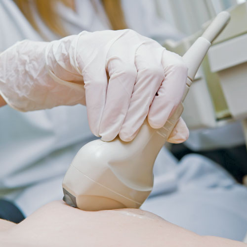
Ultrasound Imaging Santa Fe works by using a small device called a transducer, which emits high-frequency sound waves into the body. These waves bounce back off of internal organs and tissues, and are then detected by the transducer. The data collected by the transducer is then used to create images of the inside of the body, which can be viewed in real-time by medical professionals.
One of the primary benefits of ultrasound imaging is that it is non-invasive, meaning that it does not require any incisions or injections. This makes it a safer and less painful alternative to other imaging techniques, such as X-rays and CT scans. Additionally, ultrasound imaging does not use ionizing radiation, which can be harmful in large doses.
Ultrasound imaging is used in a variety of medical specialties in Santa Fe, including obstetrics and gynecology, cardiology, and radiology. In obstetrics and gynecology, ultrasound imaging is commonly used to monitor fetal development during pregnancy, as well as to diagnose and monitor conditions such as ovarian cysts and uterine fibroids. In cardiology, ultrasound imaging can be used to diagnose and monitor heart conditions, such as heart murmurs and valve abnormalities. In radiology, ultrasound imaging is used to diagnose and monitor a wide range of conditions, including liver disease, gallstones, and tumors.
What We Should Know About the Ultrasound Imaging Santa Fe:
One of the most common uses of ultrasound imaging in Santa Fe is for breast cancer screening. Breast cancer is the most common cancer in women, and early detection is key to successful treatment. Ultrasound imaging can be used in conjunction with mammography to provide a more accurate diagnosis of breast cancer. It can also be used to guide biopsies, which are used to determine whether a suspicious lump in the breast is cancerous or not.
In addition to its diagnostic uses, ultrasound imaging can also be used to treat certain medical conditions. One example is in the treatment of uterine fibroids. Uterine fibroids are non-cancerous growths in the uterus that can cause heavy bleeding and pain. In the past, the only treatment for uterine fibroids was a hysterectomy, which is the surgical removal of the uterus. However, in recent years, a new technique called ultrasound-guided focused ultrasound (USgFUS) has been developed. This technique uses ultrasound imaging to locate and destroy the fibroids using focused ultrasound waves, without the need for surgery.
Ultrasound Imaging Santa Fe How To Get it?
Another example of ultrasound imaging being used for treatment is in the field of physical therapy. Ultrasound imaging can be used to guide the application of heat or cold therapy to specific areas of the body, helping to reduce pain and inflammation. It can also be used to guide the placement of needles during acupuncture, helping to ensure that the needles are placed in the correct location.
In conclusion, ultrasound imaging is a powerful medical tool that is used extensively in Santa Fe to diagnose and treat a wide range of conditions. Its non-invasive nature, lack of ionizing radiation, and ability to produce real-time images make it an invaluable resource for medical professionals. Whether used for breast cancer screening, fetal monitoring, or physical therapy, ultrasound imaging is helping to improve patient outcomes and quality of life in Santa Fe and beyond.
Ultrasound Imaging Santa Fe How Its Work?
Ultrasound imaging, also known as sonography, is a medical imaging technique that uses high-frequency sound waves to produce images of the inside of the body. It works by using a small device called a transducer, which emits high-frequency sound waves into the body. These sound waves bounce back off of internal organs and tissues, and are then detected by the transducer.
The transducer contains crystals that vibrate rapidly when an electrical current is applied to them. These vibrations create high-frequency sound waves that travel through the body. When these sound waves encounter different types of tissues in the body, they are reflected back to the transducer.
The returning sound waves are detected by the transducer, which converts them into electrical signals. These electrical signals are then processed by a computer to create images of the internal organs and tissues of the body.
The images produced by ultrasound imaging are known as sonograms or ultrasounds. These images can be viewed in real-time, allowing medical professionals to see the movement of internal structures in the body, such as the beating of the heart or the movement of a fetus during pregnancy.
One of the key benefits of ultrasound imaging is that it is non-invasive, meaning that it does not require any incisions or injections. This makes it a safer and less painful alternative to other imaging techniques, such as X-rays and CT scans. Additionally, ultrasound imaging does not use ionizing radiation, which can be harmful in large doses.
Ultrasound Imaging Santa Fe Services:
The use of ultrasound imaging is also versatile, as it can be used for a wide range of medical applications. In Santa Fe, ultrasound imaging is used in a variety of medical specialties, including obstetrics and gynecology, cardiology, and radiology.
In obstetrics and gynecology, ultrasound imaging is used to monitor fetal development during pregnancy, as well as to diagnose and monitor conditions such as ovarian cysts and uterine fibroids. In cardiology, ultrasound imaging can be used to diagnose and monitor heart conditions, such as heart murmurs and valve abnormalities. In radiology, ultrasound imaging is used to diagnose and monitor a wide range of conditions, including liver disease, gallstones, and tumors.
Ultrasound imaging can also be used for therapeutic purposes. One example is in the treatment of uterine fibroids. Uterine fibroids are non-cancerous growths in the uterus that can cause heavy bleeding and pain. In the past, the only treatment for uterine fibroids was a hysterectomy, which is the surgical removal of the uterus. However, in recent years, a new technique called ultrasound-guided focused ultrasound (USgFUS) has been developed. This technique uses ultrasound imaging to locate and destroy the fibroids using focused ultrasound waves, without the need for surgery.

Conclusion:
In physical therapy, ultrasound imaging can be used to guide the application of heat or cold therapy to specific areas of the body, helping to reduce pain and inflammation. It can also be used to guide the placement of needles during acupuncture, helping to ensure that the needles are placed in the correct location.
In conclusion, ultrasound imaging is a powerful medical tool that uses high-frequency sound waves to produce images of the inside of the body. It is non-invasive, versatile, and does not use ionizing radiation, making it a safe and effective alternative to other imaging techniques. In Santa Fe, ultrasound imaging is used extensively in healthcare facilities to provide accurate and non-invasive diagnostic services to patients, as well as to guide therapeutic treatments.


Are you looking for the all new 2020 Nissan Maxima S 3.5L in Santa Fe?
Then Reliance Nissan is the place to visit to check out the 2020 Maxima and you can get the best deals and offers.
Visit Reliance Nissan today and learn more about 2020 Nissan Maxima S 3.5L interior, exterior, colors, engine, performance, price and more.


Hyundai i10This is another small hatchback from Hyundai, costing 4.23-5.06 lakhs, and is available in petrol and LPG variant, though the mileage of all the variants remains the same at 19.81 km/l.
The i10 comes in 5-speed manual transmission, with a displacement of 1086 cc.
Hyundai Elite i20This sedan from Hyundai costs 5.44-8.45 lakhs.
It looks, elite, like the name says, and comes in 5-speed manual transmission for all its petrol and diesel variants, with the mileage of the petrol variants at 18.6 km/l and the mileage of the diesel variants at 22.54 km/l.
Hyundai Grand i10Aptly named, this is a grand car, costing between 4.69-6.74 lakhs, and is available in petrol, diesel, and LPG variants, with the mileage of 18.9 km/l for petrol and LPG, and 24 km/l for diesel variants.
Honda Brio, Ford Figo, and Maruti Suzuki Swift are its competitors.Hyundai i20 Active This sedan costs 6.58-9.33 lakhs for its six colors.