


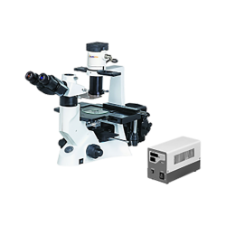
The Inverted Fluorescence Microscope is a specialized optical instrument designed for observing and analyzing fluorescent samples. Unlike a conventional upright microscope, where the objective lens is located above the specimen, the inverted microscope has its objective lens positioned beneath the sample stage. This configuration allows for the examination of thicker or larger specimens, such as cells cultured in a dish or tissue samples in a petri dish.
The key components of an inverted fluorescence microscope include:
- Illumination System: The microscope features a light source, usually a high-intensity lamp or a laser, which emits excitation light of a specific wavelength. The excitation light is directed towards the sample through a series of optical components, including filters and mirrors, which help in selecting the appropriate wavelength and focusing the light onto the specimen.
- Objective Lens: The objective lens, located beneath the sample stage, collects the emitted fluorescence from the sample. It is specifically designed to have a high numerical aperture, enabling it to gather as much light as possible from the specimen. The lens also plays a crucial role in providing high-resolution images.
- Fluorescence Filters: Fluorescence filters are positioned within the light path to selectively transmit the emitted fluorescence while blocking the excitation light. These filters ensure that only the desired wavelength of fluorescence reaches the detector, thus enhancing the contrast and specificity of the observed fluorescence signal.
- Dichroic Mirror: A dichroic mirror, also known as a beamsplitter, is used to separate the emitted fluorescence from the excitation light. It reflects the fluorescence signal towards the detector while allowing the excitation light to pass through or reflect away.
- Detector: The detector, typically a high-sensitivity camera or a photomultiplier tube (PMT), captures the emitted fluorescence and converts it into an electronic signal. The camera records the fluorescence signal as an image, while a PMT measures the intensity of the fluorescence for quantitative analysis.
- Sample Stage: The sample stage is located above the objective lens and provides a stable platform to hold the specimen. It is adjustable in height, allowing precise focusing of the specimen. Some advanced inverted microscopes may incorporate environmental control elements, such as temperature and CO2 regulation, to maintain the viability of live samples during long-term observations.
In summary, an inverted fluorescence microscope offers a versatile and powerful tool for studying fluorescence-labeled samples. Its unique design allows for the examination of thicker specimens and provides valuable insights into various biological processes, such as cellular interactions, protein localization, and molecular dynamics. For fore visit www.Labozon.com


