


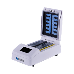
The FISH (Fluorescence In Situ Hybridization) Hybridization System is a sophisticated molecular biology technique used to visualize and detect specific DNA or RNA sequences within cells or tissues. It combines the principles of nucleic acid hybridization with fluorescence microscopy, allowing researchers to precisely locate and quantify target sequences with high sensitivity and specificity.
The FISH Hybridization System typically involves the following steps:
- Probe Design: Specific DNA or RNA probes are designed complementary to the target sequence of interest. These probes are typically labeled with fluorescent molecules, such as fluorophores or chromophores, which emit light when excited by a specific wavelength.
- Sample Preparation: Cells or tissue samples are fixed onto a microscope slide and permeabilized to allow the probes to penetrate and hybridize with the target nucleic acid sequences.
- Hybridization: The labeled probes are applied to the sample and allowed to hybridize with the complementary target sequences. This step is carried out under controlled conditions to ensure optimal probe-target binding.
- Washing: Unbound probes are removed by washing the sample with a series of buffer solutions, minimizing background fluorescence and enhancing signal-to-noise ratio.
- Visualization: The sample is examined under a fluorescence microscope equipped with appropriate filters to selectively excite the fluorophores attached to the hybridized probes. This results in the visualization of fluorescent signals localized to the specific target sequences within the sample.
- Image Analysis: Fluorescent signals are captured using digital imaging systems and analyzed using specialized software to quantify signal intensity, determine subcellular localization, and assess spatial distribution of the target sequences.
The FISH Hybridization System has a wide range of applications in molecular biology, genetics, cytogenetics, and clinical diagnostics. It is commonly used for gene mapping, chromosome analysis, detection of chromosomal abnormalities, identification of microbial pathogens, and gene expression analysis in various biological samples.
By enabling precise and specific detection of nucleic acid sequences within cells and tissues, the FISH Hybridization System has revolutionized our understanding of cellular processes, disease mechanisms, and genetic variation, making it an invaluable tool in biological research and medical diagnostics.


Global flourescent in situ hybridization probe market is expected to register a substantial CAGR of 6.6% in the forecast period of 2019-2026.
Market Definition: Global Flourescent in Situ Hybridization (FISH) Probe Market Fluorescent in situ hybridization (fish) would help in the identification of position of genes.
It is used in clinic for diagnosing the chromosomal abnormalities including duplications, deletions and translocations etc.
FISH has wide applications in research labs and clinics for monitoring the after effects of therapies, tracking of cells after transplantation etc.
Many developments are taking place in FISH market including DNA probe-based diagnostics which assists in the determination of suspected diseases caused by any bacteria.
Get Sample Report: https://databridgemarketresearch.com/request-a-sample/?dbmr=global-flourescent-in-situ-hybridization-fish-probe-market Major Market Competitors/Players Few of the major market competitors currently working in the global flourescent in situ hybridization probe market are Oxford Gene Technology, Agilent Technologies Inc, PerkinElmer Inc,, Horizon Discovery Group plc, BioDot, Sysmex Singapore Pte.


Global “Fluorescence In Situ Hybridization (FISH) Market” report offers an overview of the market size, share, trends, drivers, and barriers with respect to the Fluorescence In Situ Hybridization (FISH) Industry.
The competitive data type analysis includes capacity, market share, profit margin, market growth, consumer consumption, imports, exports, revenue, and etc.
The global Fluorescence in Situ Hybridization (FISH) Imaging Systems Market is estimated to touch US$ 1.4 billion by the completion of the prediction period.
Increasing occurrence of target sicknesses tips to medical earnestness for acceptance of speedy analytical substitutes for example Fluorescence in Situ Hybridization (FISH) Imaging Systems, this is expected to trigger the demand in the upcoming years.
Furthermore, increasing alertness about the related profits of this system, for example speedy investigation, improved imagining, greater exactness of results, and an extensive variety of use, is too between the most important issues accountable for amplified acceptance.
The Fluorescence in Situ Hybridization (FISH) Imaging Systems market on the source of Type of Application could span Hereditary Sickness Analysis, Cancer Analysis [Breast Cancer, Lung Cancer and Others].